Planctoteuthis danae
Brief diagnosis:
A Planctoteuthis with:- 12-13 suckers on proximal third of arm IV (distal two thirds bare).
- Fin length more than 50% of mantle length.
- Arms
- All arms weakly muscled (especially arms IV) relative to the well muscled tentacles.
- All arms with attenuate tips; arms I-III with many very small suckers on tips.
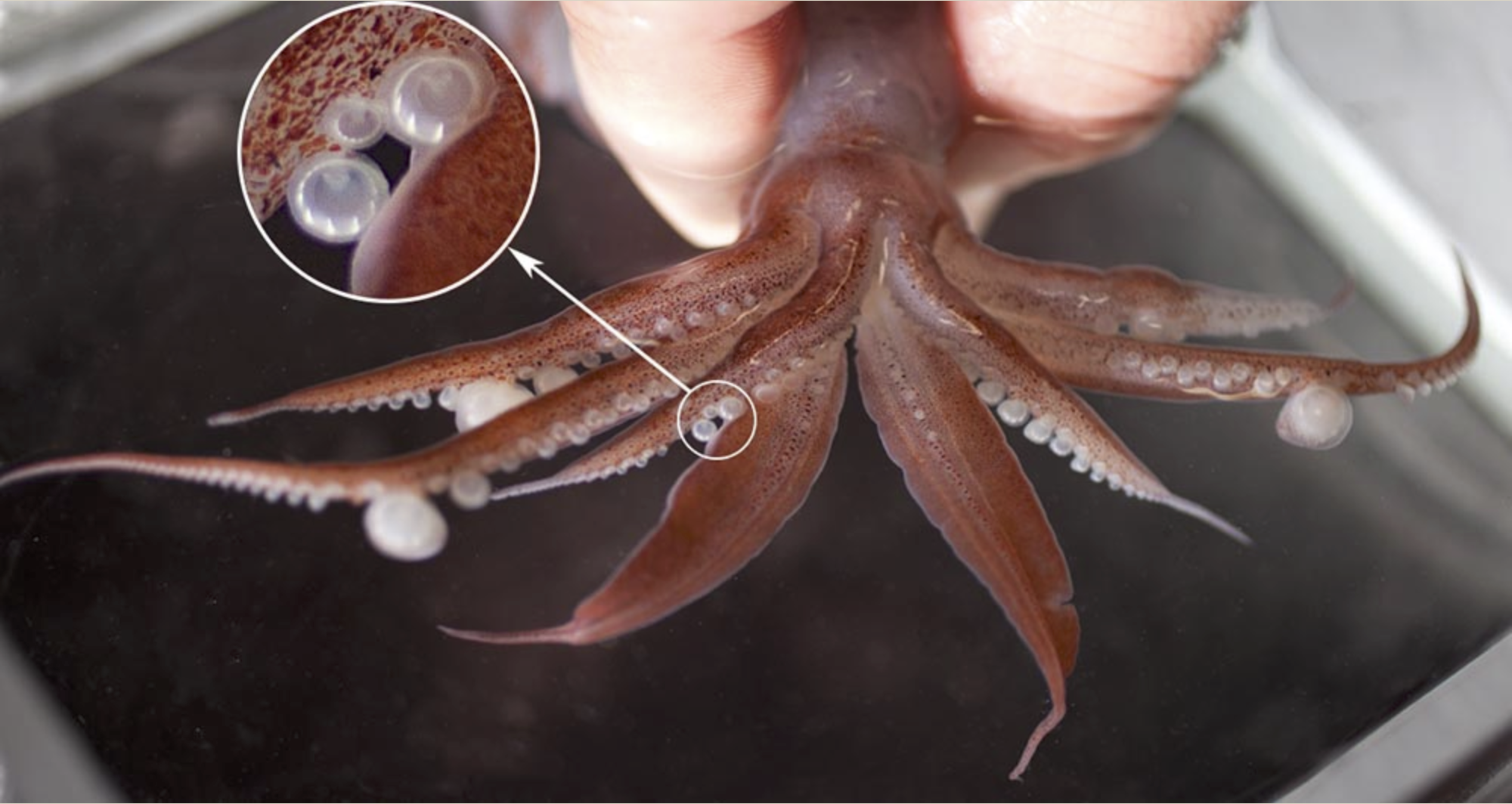
- Large females with arms II and III each with two greatly enlarged suckers (one of which is much larger than the other) in mid-arm.
- Mid-arm I suckers of ventral series much larger than those of dorsal series, but not nearly as large as enlarged suckers of arms II and III.
- Arms IV each with 12-13 suckers in proximal 1/3 of arm, aligning in virtually a single series; distal 2/3 of arm without suckers.

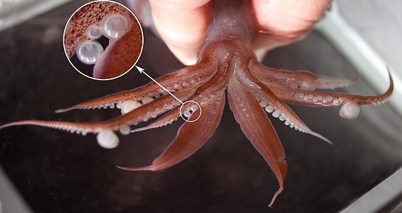

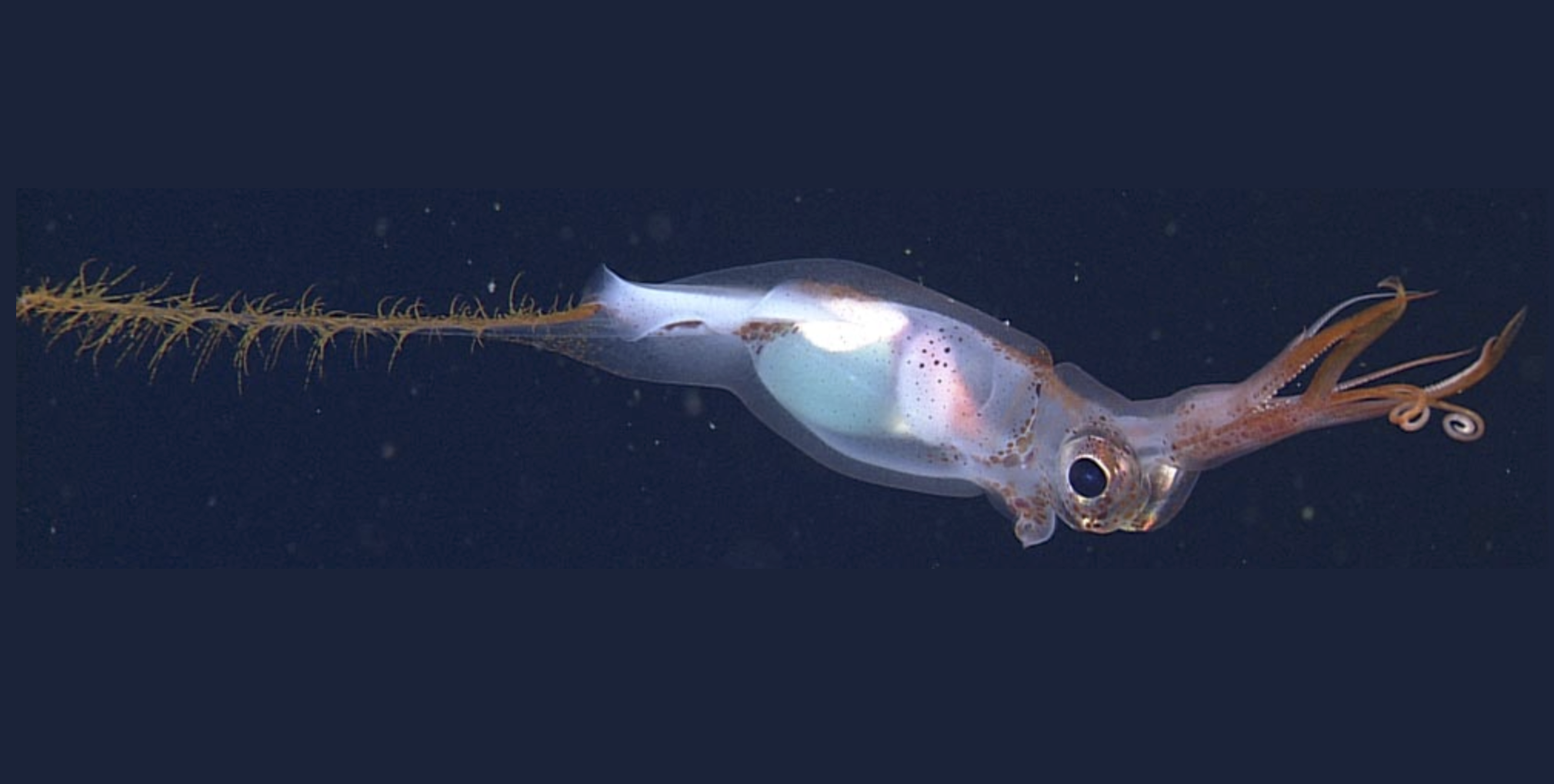
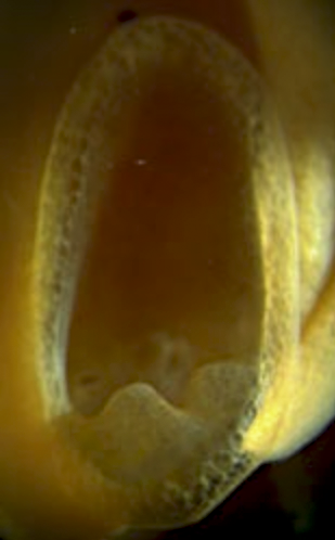
Figure. Anterodorsal view of a live, nearly mature female P. danae, 130 mm ML, showing arm crown. Insert shows enlargement of suckers of arm I. Note the larger size and larger teeth of the suckers of the ventral series. Photograph by Karen Osborn. © MBARI.
- Tentacular clubs
- Clubs bilaterally symmetrical and lack keels.
- Clubs bilaterally symmetrical and lack keels.
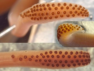

Figure. Oral views of the clubs and club suckers of P. danae. Left - Three views of the club, holotype, ca. 65 mm ML. Photographs by R. Young. Right - View of the club and basal club sucker (left) and mid-club sucker, 47 mm ML, showing differences in aperature width. Note that this difference is apparent in the lower photograph at left. Drawings from Roper and Young (1967).
- Head
- Beaks: Descriptions can be found here: Lower beak; upper beak.
- Beaks: Descriptions can be found here: Lower beak; upper beak.
- Funnel
- Posteriorly located antitragus of funnel locking-apparatus with two distinct lobes.
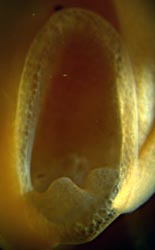
Figure.
 Frontal view of the funnel locking-apparatus, P. danae, subadult, eastern Pacific. Photograph by R. Young.
- Fins
- Fin length 52% of ML.


Comments
This species was originally placed in the genus Valbyteuthis Joubin, 1931, which was subsequently synonomized with Planctoteuthis (Young, 1991).
Planctoteuthis danae: Additional morphology.
Richard E. Young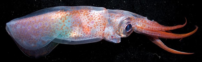
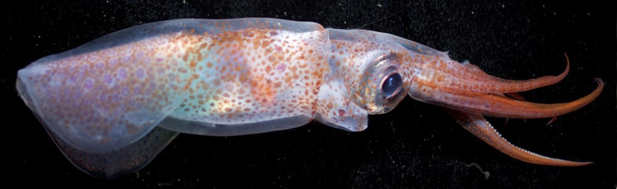 Figure. Side view of a mature male of P. danae, 92 mm ML, in a shipboard aquarium. Note the thick gelatinous layer over the mantle, tail, head and arm bases. This layer is, presumably, vacuolated and aids buoyancy. Photograph by Karen Osborn. © MBARI
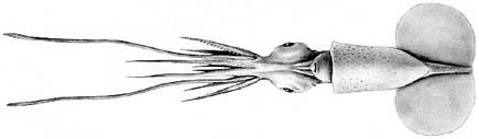
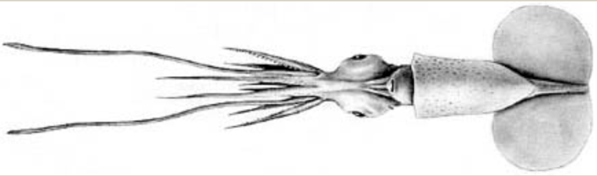 Figure. Ventral view of P. danae, 47 mm ML. Drawing from Roper and Young (1967).
-
- Arm lengths: arms I: 38-44% of ML (subadults); arms III: 50-55% of ML (subadults); arms IV: 74-91% of ML (subadults).
- Arm I with ca. 40 pairs of suckers; arm II with 45 pairs of suckers; arm III with 35 pairs of suckers; arm IV with 12 suckers on proximal quarter (Joubin, 1931).
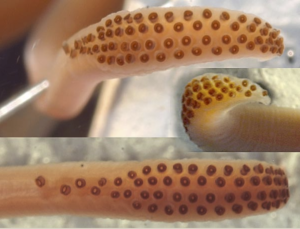
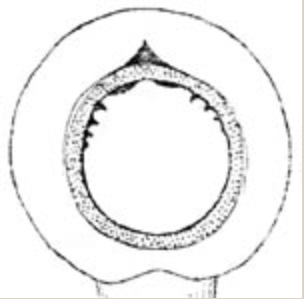
- Largest arm suckers in small squid with 7-9 small, blunt teeth on distal margin of ring; proximally teeth fuse into a virtually smooth ring.
- Lateral arms in holotype with two greatly enlarged (2-3 times larger) suckers; enlarged suckers at position of sucker pairs 15 and 16 on arms II and 18 and 19 on arms III (Joubin, 1931).Arms.
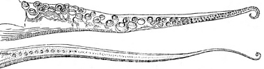
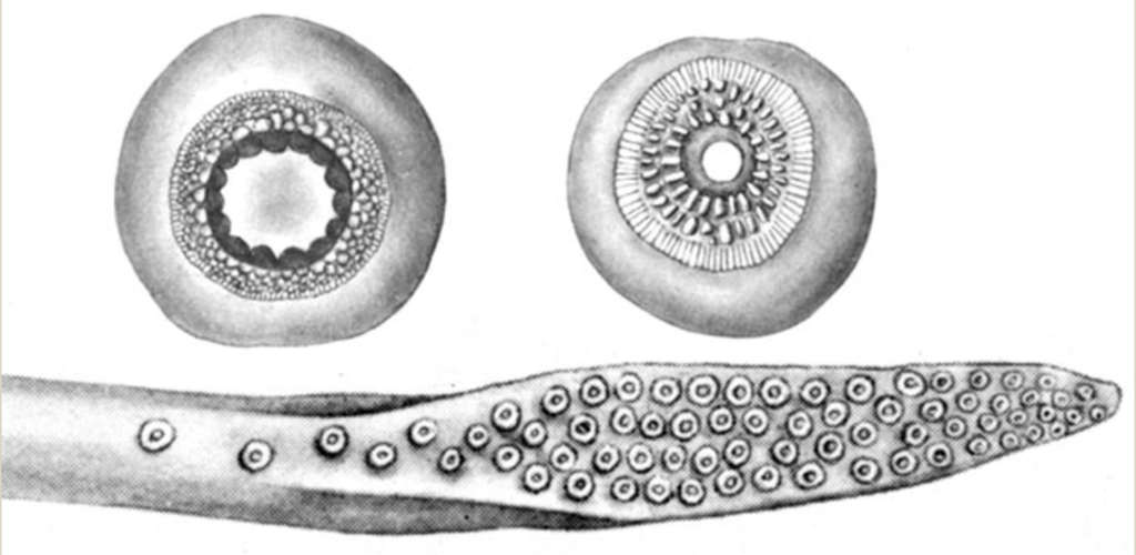
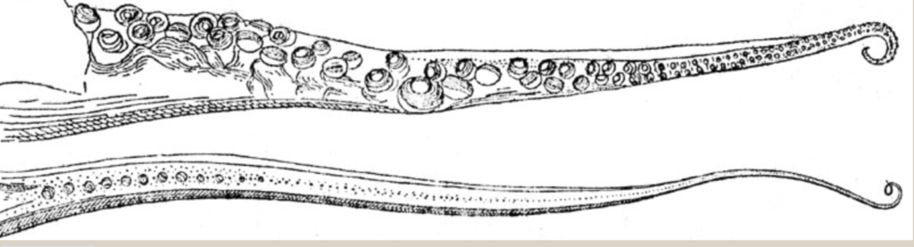 Figure. Oral views of arm III (top) and arm IV of P. danae, holotype, ca 65 mm ML. Drawings from Joubin (1931).
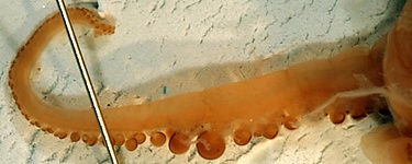
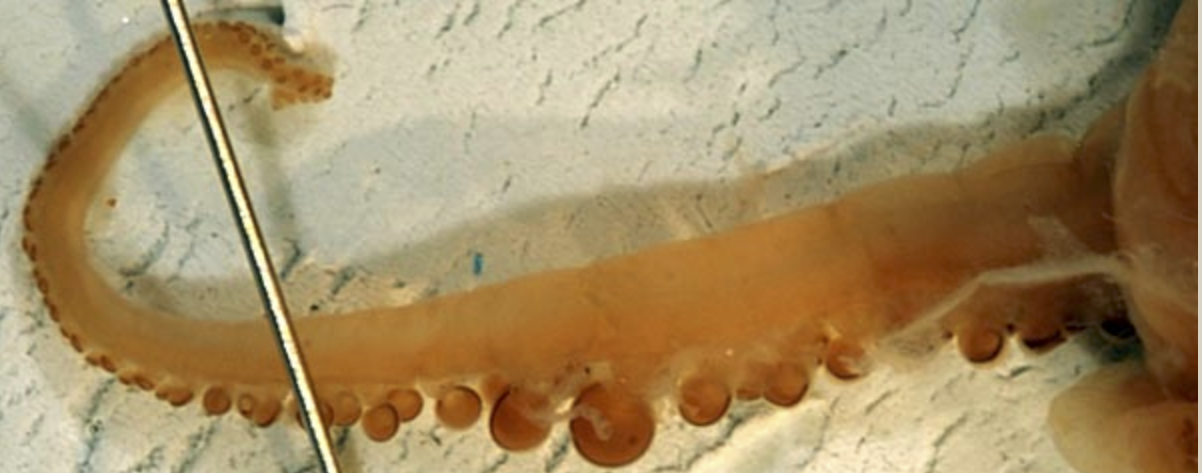
 Figure. Side view of arm III of P. danae, holotype, ca. 65 mm ML, showing two suckers with initial enlargement in mid-arm. Photograph by R. Young.
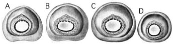
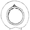
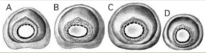
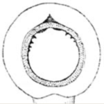 Figure. Left - Oral views of large arm suckers of P. danae, 47 mm ML, eastern tropical Pacific Ocean. A-D - Suckers from arms I-IV respectively. Drawings from Roper and Young (1967). Right - Oral view of early stage of enlargement of sucker of a lateral arm, holotype. Drawing from Joubin (1931).
- Tentacular clubs
- Club of holotype with about 60 suckers (Joubin, 1931).
- Club length 12-18% of ML.
- "Carpal" suckers on club with broader apertures than regular club suckers.
- Viscera
- Reproductive structures

 .
. 
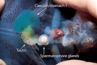
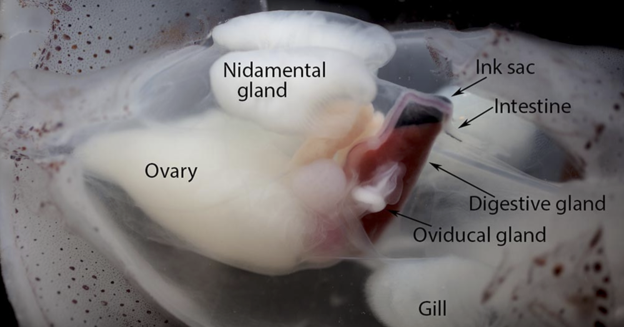
 Views of the viscera of P. danae. Top - Figure. Ventrolateral view of the nearly mature female, 130 mm ML. Photograph by uncertain. Bottom - Ventral view of the just mature Male, 92 mm ML. Photograph by Karen Osborn. © MBARI.
- Reproductive structures

Comments
This description is from Roper and Young (1967) unless otherwise indicated.Paralarvae/juveniles:
Figure.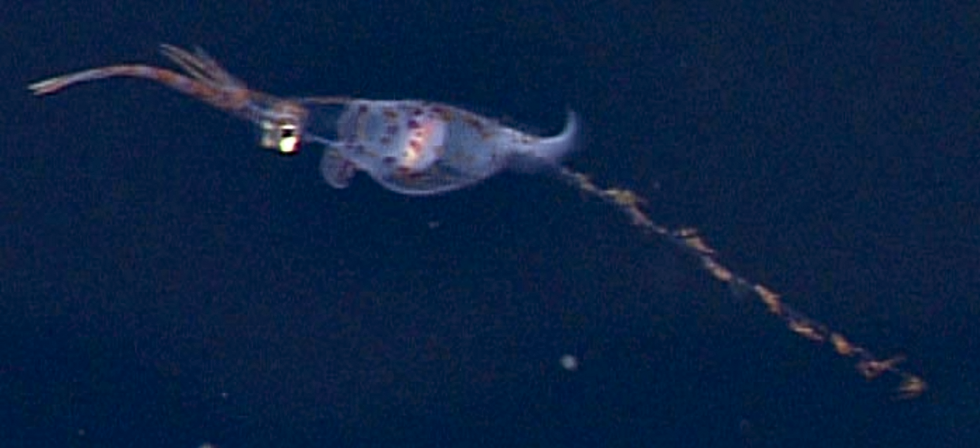 Side view of a very young P. danae, 20 mm ML (About the size of the largest drawing below).
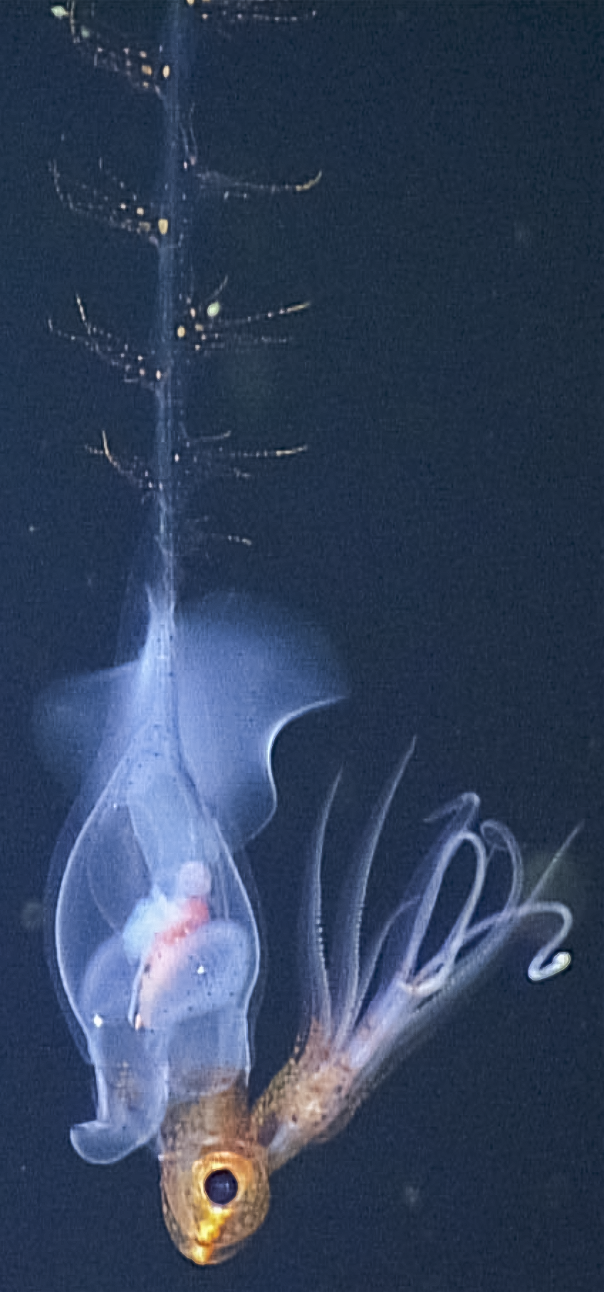
In situ ROV photograph taken at a depth of 829 m. Note long neck and eyes strongly protruding ventrally.© 2013 MBARI.
Paralarvae of P. danae are most easily distinguished from other doratopsis paralarvae in Hawaiian waters by:
Side view of a very young P. danae, 20 mm ML (About the size of the largest drawing below).
In situ ROV photograph taken at a depth of 829 m. Note long neck and eyes strongly protruding ventrally.© 2013 MBARI.
Paralarvae of P. danae are most easily distinguished from other doratopsis paralarvae in Hawaiian waters by:
- The dorsal position of the oesophagus in the brachial pillar (see figure).
- The small number of brachial pillar chambers (two).
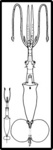
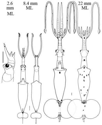 Figure.
 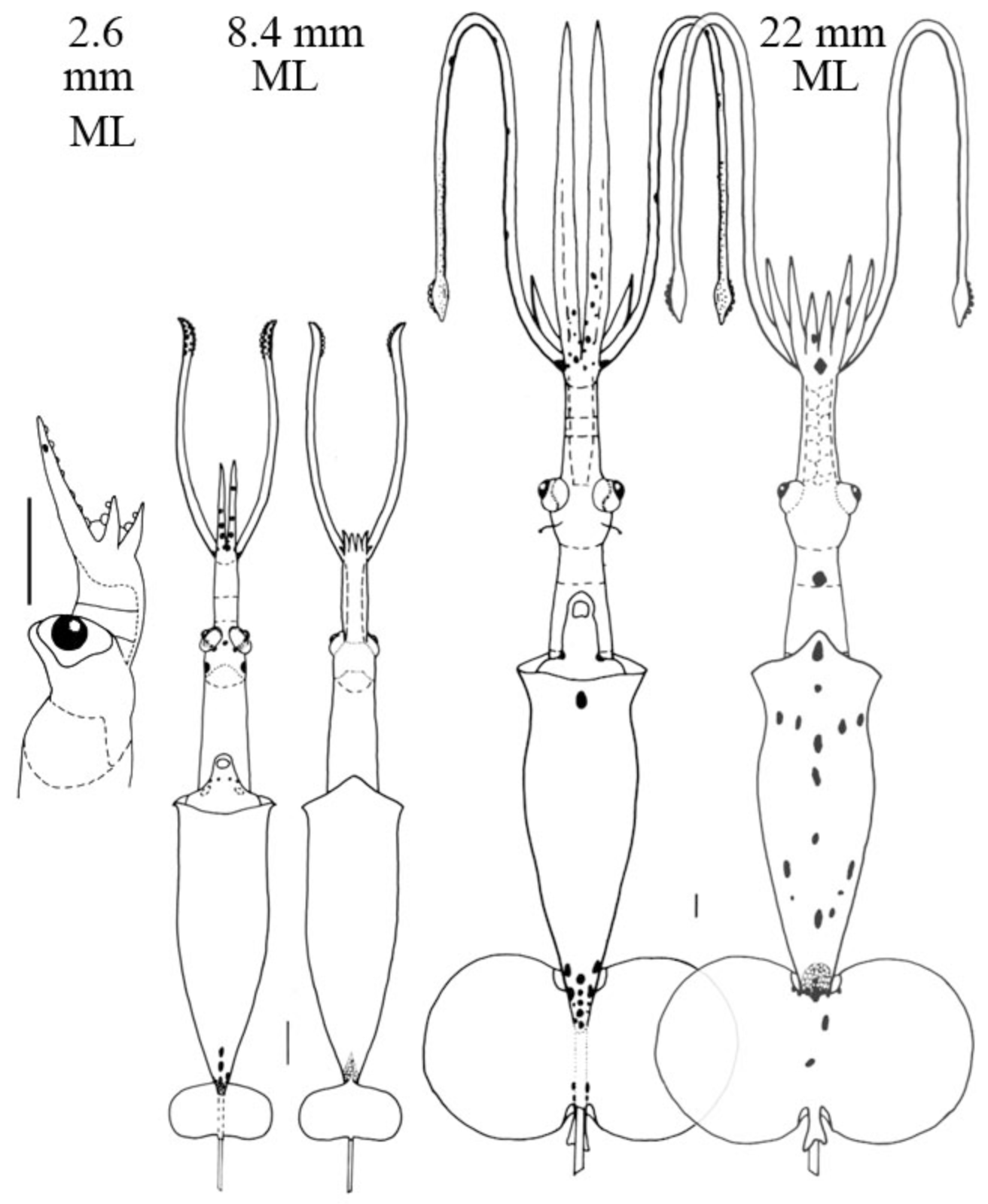 Paralarvae of Planctoteuthis danae from the waters off Hawaii: Thumbnail - Ventral view of the two paralarvae showing relative sizes. Left - Side view of the head, 2.6 mm ML, showing the ventrally protruding eye, the dorsal position of the esophagus and the small number of chambers in the brachial pillar. Middle - Ventral and dorsal views, 8.4 mm ML. Right - Ventral and dorsal views, 22 mm ML. Scale bars are 1 mm. Drawings from Young, 1991. Note the very elongate arms IV in the 22 mm ML paralarva which is typical of the doratopsis stage in the family. Compare this with the relative arms length of the near-adult female below.
Adults:
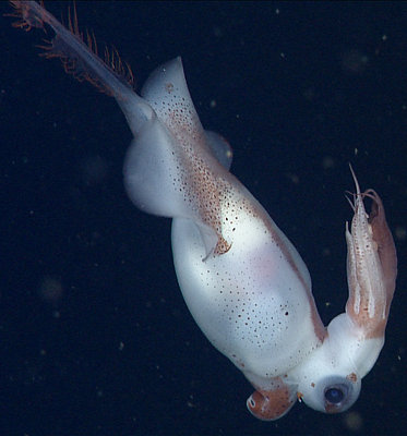
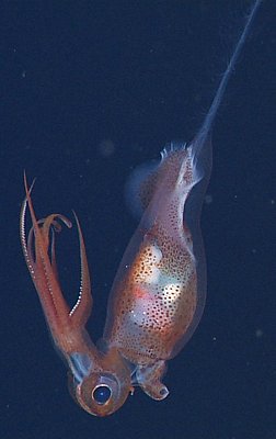
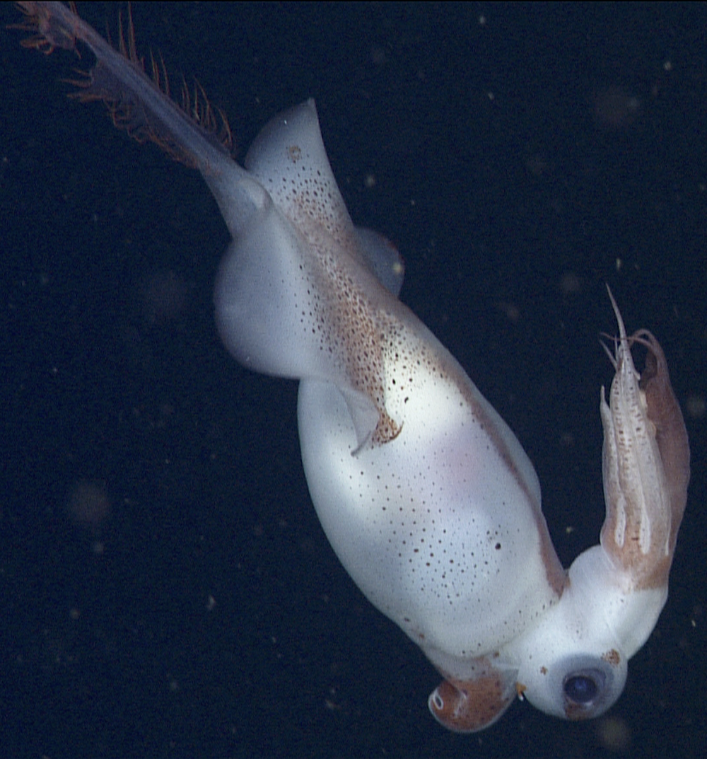
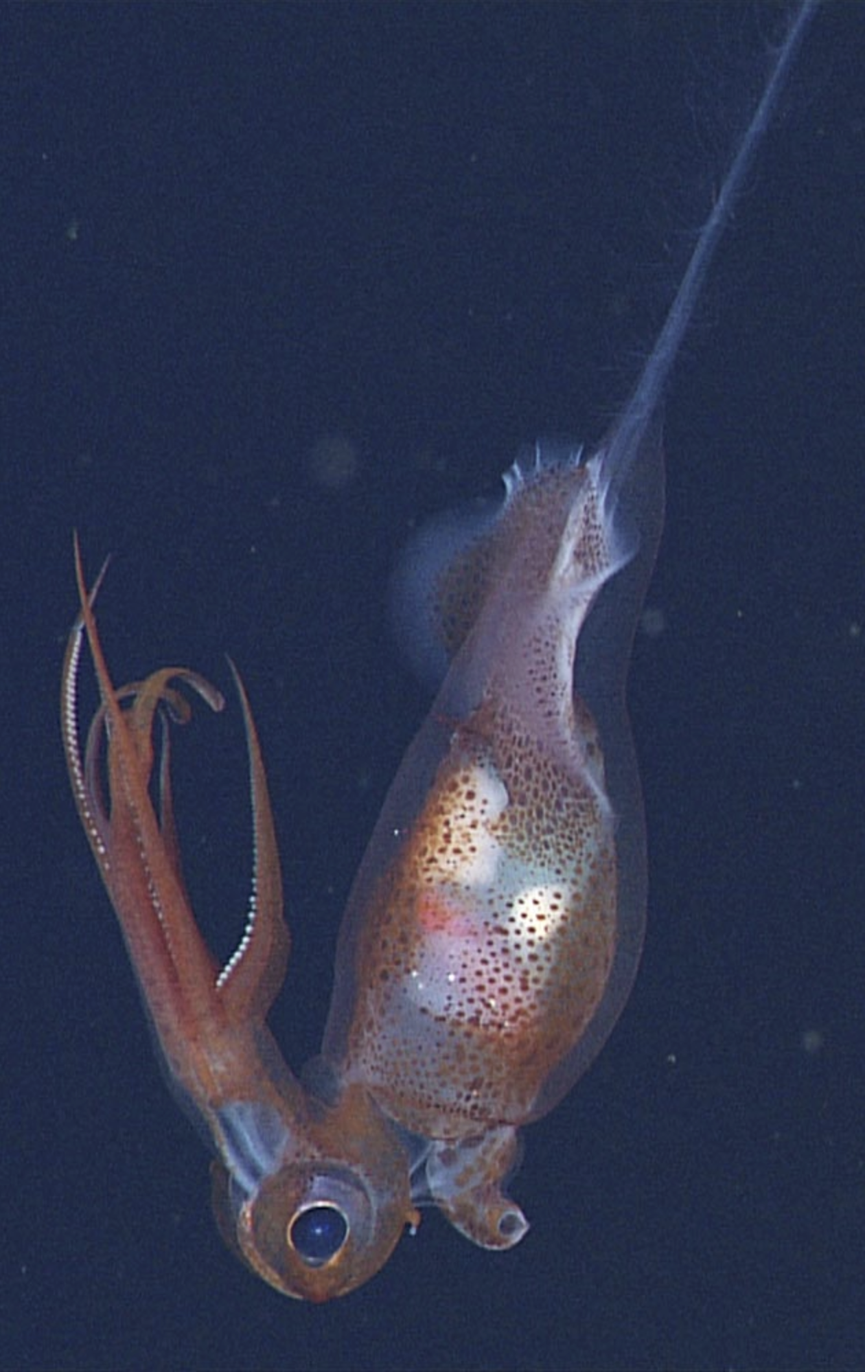 Figure. Side views of P. danae. Left - near-mature female, pictured earlier on this page, at a depth of 1441 m in the Gulf of California. Right - A presumed mature male, judging from the large white structure (probably the spermatophore gland complex) on the ventral side of the viscera, at a depth of 1193 m. This male appears to be slightly larger and more mature than the one captured and seen on the adjoining page. Note that the arms IV are about the same length as arms III in both squid. Both photographs © 2013 MBARI.
Clarke, M. R. and C. C. Lu. 1975. Verical distribution of cephalopods at 18 N 25 W in the North Atlantic. Journal of the Marine Biological Association of the United Kingdom, 55 (1): 165-182.
Joubin, L. 1931. Notes preliminaires sur les cephalopodes des croiseires du “Dana” (1921-1922). Annales de l’Institut Oceanographique, 10: 169-211.
Lu, C. C. and M. R. Clarke, 1975. Vertical distribution of cephalopods at 11 N 20 W in the North Atlantic. Journal of the Marine Biological Association of the United Kingdom, 55 (2): 369-389.
Nesis, K. N. 1982. Abridged key to the cephalopod mollusks of the world's ocean. 385+ii pp. Light and Food Industry Publishing House, Moscow. (In Russian.). Translated into English by B. S. Levitov, ed. by L. A. Burgess (1987), Cephalopods of the world. T. F. H. Publications, Neptune City, NJ, 351pp.
Roper, C. F. E. and R. E. Young (1967). A review of the Valbyteuthidae and an evaluation of its relationship with the Chiroteuthidae. Proc. U.S. Nat. Mus., 123: 1-9