Asperoteuthis mangoldae
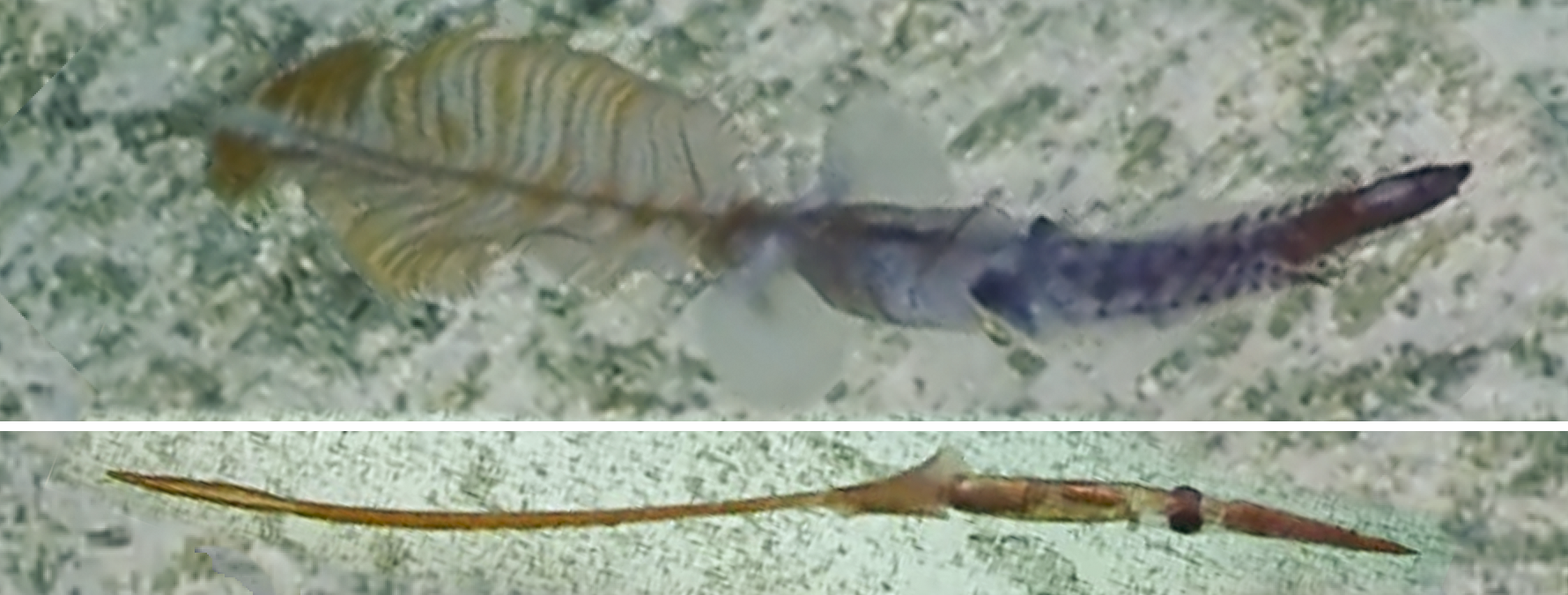
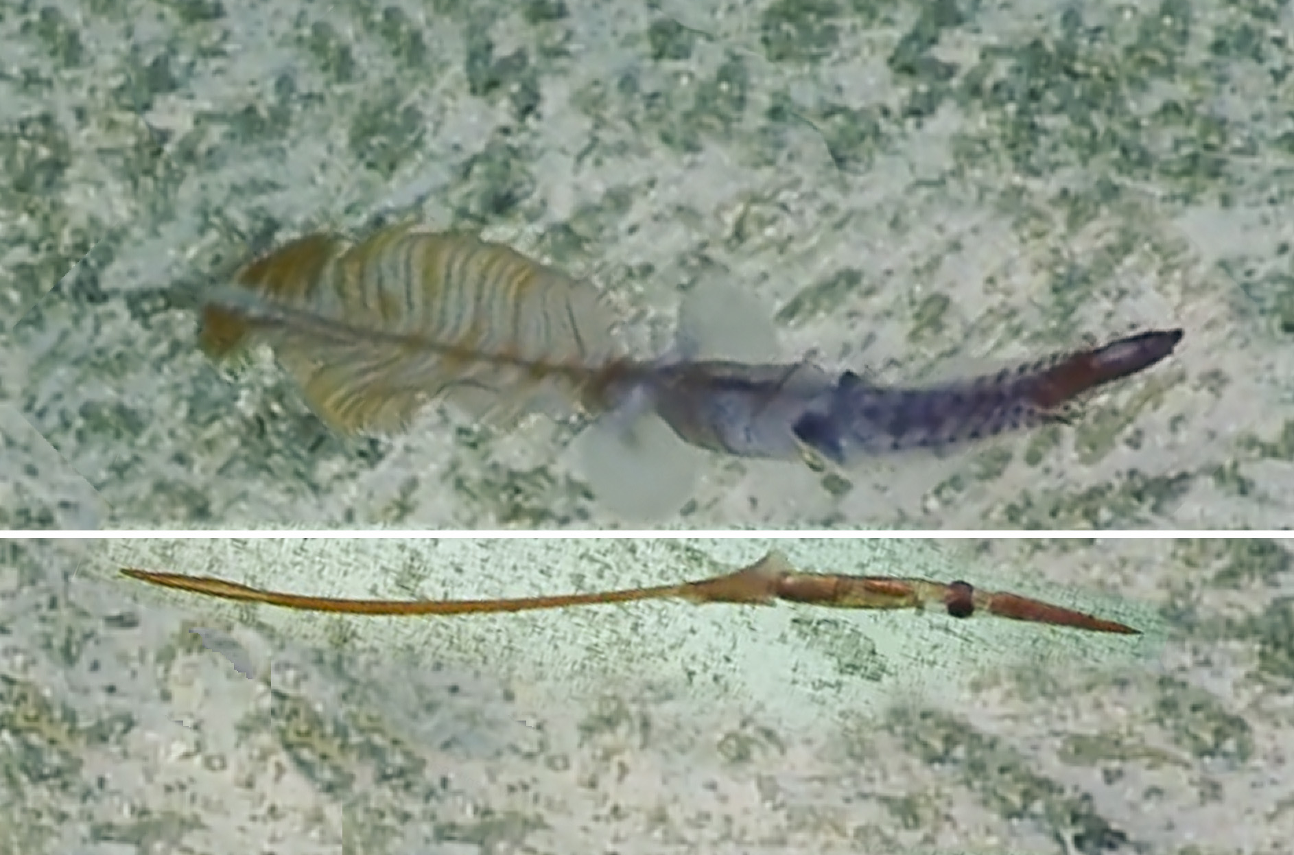
Asperoteuthis mangoldae, 930m, Jarvis Seamount, filmed by EV Nautilus
 The upper member of the featured images above, shows the expanded secondary fin(s) of the tail (the tail may have two sets of secondary fins (as suggested by this image) with the smaller, darker one at the posterior end of the tail) with a striped, elliptical leaf-shaped appearance. This squid was observed swimming close to the ocean floor at 930 m depth on the Jarvis Seamount (at the equator about 160º West), from an ROV operated by the E/V (Exploration Vessel) Nautilus. The squid was not captured. The lower video clip shows (to the surprise of everyone involved at the time (i.e., July 2019)) that the secondary fins could mostly disappear (in about 10 seconds) by collapsing onto the extended rod-like gladius of the tail; the complete collapse takes a bit longer, the lower image was photographed about 1 minute after the upper image. The video can be seen here. Prior to this short video of the living squid, our knowledge Asperoteuthis mangoldae was based on the examination of of a limited number of preserved specimens. It appears to be the smallest member of the genus (a mature male is known with a ML of 100 mm; no mature females have been found). The species is very fragile, with a gelatinous consistency similar to that of Grimalditeuthis bonplandi. A. mangoldae is very different from other members of the genus, especially in features of the tentacle club, funnel-mantle locking apparatus and absence of skin tubercules (see the Asperoteuthis page for a comparison between the three species).
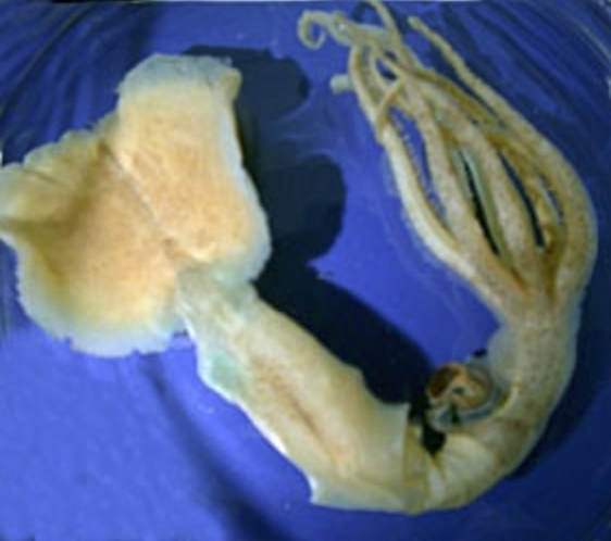
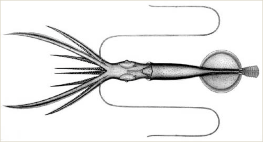 Figure 2. A. mangoldae. Left: Dorsal view, paratype, 100, mm ML, mature male, preserved, © R. Young. Right: Holotype (NMNH 729749), ventral view, 80 mm ML, immature male, drawing by A. Hart, © C. F. E. Roper.
- Arms
- Largest arm suckers with 6-10 truncated teeth on distal half of ring.
- Arms without enlarged suckers; largest suckers in proximal half of arm with sucker size slowly decreasing distally; arms I-III with suckers of comparable size, arms IV with distinctively smaller suckers.
- Tentacles
- Suckers with ca. 8 large, truncated teeth on distal half of inner ring grading to ca. 17 small, truncated teeth on proximal half.
- Club with very broad proximal protective membrane and long terminal papilla.
 .
.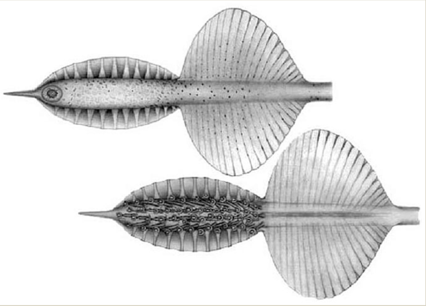
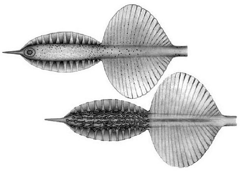
Figure 3. A. mangoldae, holotype. Aboral (top) and oral (bottom) views of the tentacular club. Drawing by A. Hart, © C. Roper.
Very few A. mangoldae have been captured with the clubs attached as the long, thin tentacles are easily broken off during capture. With tentacles missing, this species can be confused with Grimalditeuthis bomplandi in its general shape and consistency. However, many differences easily separate them including presence of the funnel-mantle fusion and three large papillae on each arm-sucker base in G. bomplandi.
- Head
- Beaks: Descriptions can be found here: Lower beak; upper beak.
- Beaks: Descriptions can be found here: Lower beak; upper beak.
- Funnel
- funnel-locking apparatus oval with a rounded ridge in the midline (tragus?) and an elongate, deep, curving groove laterally; mantle locking component with matching curved ridge. Antitragus absent.
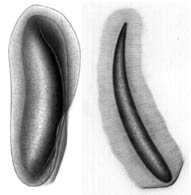
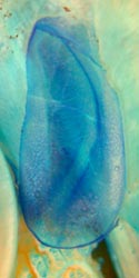
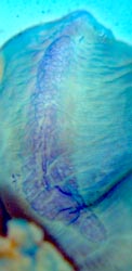
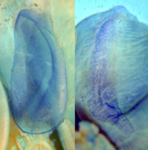
Figure 4. Frontal views of the funnel-mantle locking apparatus of A. mangoldae. Funnel (left) and mantle (right) components, 100 mm ML, mature male, paratype, stained with methylene blue. Photographs by R. Young.
- funnel-locking apparatus oval with a rounded ridge in the midline (tragus?) and an elongate, deep, curving groove laterally; mantle locking component with matching curved ridge. Antitragus absent.
- Mantle
- Mantle and other integument without tubercles.
- Mantle and other integument without tubercles.
- Primary Fins
- Fin length 40% of ML.
- Fin width approximately equals fin length.
- Photophores
- Club-tip photophore small, terminal papilla large.
- Small embedded photophores present on aboral surface of club.
- Ventral eyeball photophore present.
- Luminescent pads present on tentacle stalks.

Comments
More details of the description can be found here.
A. mangoldae possesses a large secondary tail but a specimen with an attached tail has not been captured.
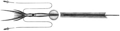
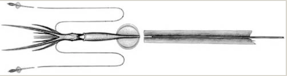 Figure 5. The drawing is a reconstruction based on the collection of a damaged tail in the same midwater tow with a small (80 mm ML) A. mangoldae. The tail did not match the broken end of the gladius from the specimen indicating that a piece of the tail was missing. Tentacular clubs, missing from specimen, were added to drawing. Drawing by A. Hart.
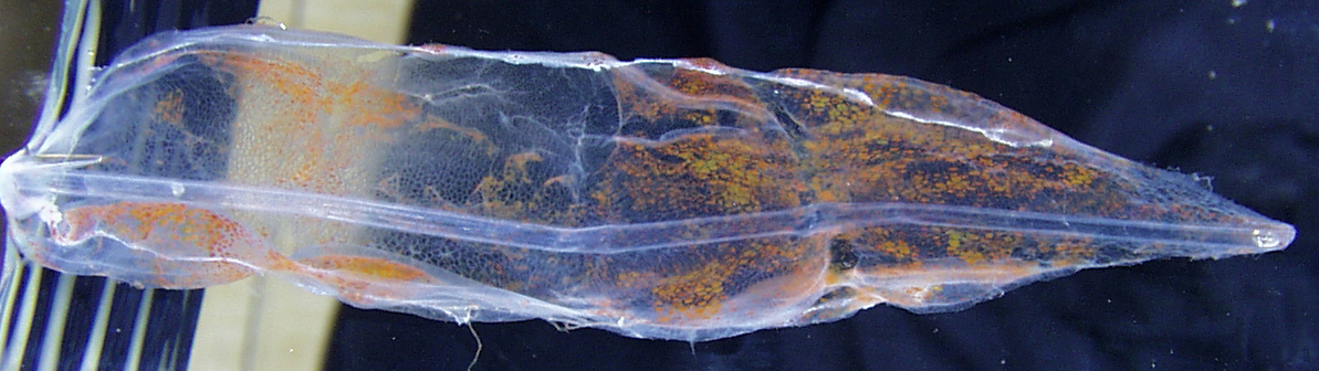
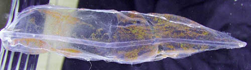
Figure. The photograph shows the expanded secondary fin of the tail of a large Asperoteuthis from the same Hawaiian locality as in Fig. 5 and possibly belonging to a large A. mangoldae although the shape differs greatly from that of the small specimen above; however it looks similar to that in the featured image.
In addition to the features listed in the Diagnosis, A. mangoldae differs from A. acanthoderma in (1) the weaker consistency of the arms and mantle, (2) the dentition of the arm (6-10 vs 3-4) and club (25 vs 9 teeth) suckers, (3) the smaller and more circular terminal club photophore, (4) the longer terminal papilla on the club and (5) the broader fins (width ca 115% of length vs ca 75% of length). In addition, the club suckers of A. acanthoderma are on shorter stalks, the sucker rings are more elongated in the oral-aboal directiion and suckers of arms [[ This paragraph should be in the Asperoteuthis page ]] FIX
Vertical distribution.
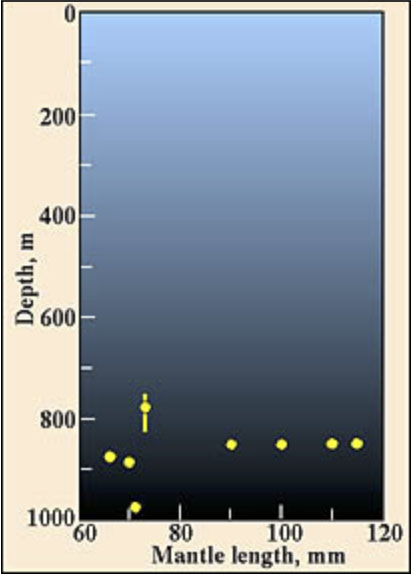
Off Hawaii only eight specimens were captured during this study. All were captured during the day at depths between 775 and 975 m. The absence of captures in the heavily sampled waters from 0-400m depth at night suggests that this species is a deep living, non-vertical migrator. Geographical distribution. Type locality: Hawaiian waters at 21°25'N, 158°20.5'W. The holotype was captured in an opening-closing trawl between 820 and 870 m depth during the day (1411-1616 hrs). This species is presently known only from the waters off the Hawaiian Islands.
- Largest arm suckers with ca. 9 truncated teeth on distal half of ring.

 Figure. Arm suckers of A. mangoldae. Top left - Distal view of a large arm III sucker, 100 mm ML, mature male, preserved. Photograph by R. Young. Top right - Oral view of one arm sucker (sucker 10, arm II, holotype), scanning electron micrograph (SEM). A piece of debris lies inside the sucker acetabulum. Bottom - Oblique view of the same arm II sucker, SEM. Note that the teeth project vertically outward from the face of the sucker. Photographs by R. Young.
 - Arms IV longest but relative size decreases with increasing squid size.
- Arm IV suckers much smaller than arm III suckers.
 Figure. Oral views of arms III (top) and IV (bottom) of A. mangoldae at the same distance from the arm bases, 100 mm ML, mature male, preserved. Photograph by R. Young.
 - Sucker stalks with broad bases
- Largest arm suckers with ca. 9 truncated teeth on distal half of ring.

 Figure. Arm III suckers of A. mangoldae. Left - Oral view of three suckers and their stalks, NMNH 729752. Drawing by A. Hart. Right - Dorsal view of mid-arm suckers, 100 mm ML, mature male, preserved. Photograph by R. Young.
- Tentacles
- Suckers with ca. 8 large, truncated teeth on distal half of inner ring grading to ca. 17 small, truncated teeth on proximal half.

 Figure. Oral views from different angles of two large club suckers from A. mangoldae, holotype, scanning electron micrographs. Note that the teeth project vertically outward from the face of the sucker. Photographs by R. Young.
 - The club sucker stalks consists of a single portion.
- Stalks from the medial and lateral series approxmately the same length.

 Figure. Oral views of club suckers and stalks of A. mangoldae. Left - A pair of club suckers with stalks from the medial and lateral sucker series, NMNH 729749, holotype. Flag on lateral stalk was not always present. Drawing by A. Hart. Right - Most of the distal portion of the club, holotype, preserved. Photograph by R. Young.
 - Club with 13 trabeculae in each distal protective membrane; 24-25 trabeculae in each proximal protective membrane.
- Suckers with ca. 8 large, truncated teeth on distal half of inner ring grading to ca. 17 small, truncated teeth on proximal half.
- Head
- Head elongate with long neck and brachial pillar.
- Olfactory organ on long stalks and postioned well posterior to eyes at level of posterior end of cephalic cartilage.
- Radula
- Radula with strong secondary cusps on both the rhachidian and first lateral teeth.
- Beaks
- Upper beak with strongly recessed shoulder with sharp ridge on oral and aboral sides of shoulder. Oral ridge extends well onto pallet. Ridges absent from aboral surface of pallet. Rostrum short and curved at tip.
- Lower beak with wing fold obscuring beak angle and extending nearly to tip of rostrum. Lateral wall with weak oblique fold.
- Funnel
- Funnel valve present.
 Figure. Ventral view of and funnel organ of A. mangoldae, NMNH 729752, with the funnel wall cut and spread open. Drawing by A. Hart.
- Funnel valve present.
- Mantle
- Mantle tissue with gelatinous consistency and filled with small vesicles.
- Mantle musculature terminates in anterior third of fins.
- Fins
- Combined fin width exceeds fin length.
- Photophores
- Terminal aboral club photophore small.
- Small embedded photophores on aboral surface of club laterally near protective membranes. About 8 in distal section of club on each side and 5 in proximal section on each side.

 Figure. Aboral view of the club of A. mangoldae. Top - NMNH 729749, holotype. Drawing by A. Hart. Bottom - Distal portion of club, holotype, preserved. Photograph by R. Young.
 - Eye photophore
- Tentacles

 Figure. The eyeball of A. mangoldae. Left - Ventral view, USNM 729752, showing large photophore. Drawing by A. Hart. Right - Side view of a damaged head and eye. The iridescent tissue has pulled free of the eyeball and the lower portion, appearing dark red, is the photophore that was originally positioned on the ventral surface of the eyeball, paratype, 100 mm ML, mature male, preserved. Photograph by R. Young.
- Visceral photophores
- Visceral photophores absent.
- Visceral photophores
 Figure. Ventral view of the intestine of A. mangoldae, NMNH 729752. Above the intestine, in the drawing, is the ink sac and below the intestine, in the drawing, is the cephalic vein.
- Pigmentation
- Pigmentation in typical chromatophores; chromatophores scattered on body and buccal membrane.
- Viscera
- Penis long and slender in mature male.
 Figure. Ventral view of the mantle cavity and funnel of A. mangoldae, paratype, 100 mm ML, mature male, preserved. Note the long, slender penis extending from the gill base past the blue-stained funnel locking-apparatus and carrying a lobate tip.
- Penis long and slender in mature male.
- Measurements
Holotype NMNH 729749 Clarke 70-12-31 Clarke 70-12-31 Clarke 70-12-31 Paratype SBMNH 369535 Sex Imm. male -- -- -- Mature male Mantle length 80 128 120 82 100 Fin length 30 56 55 34 53 Fin width 37 72 66 39 67 Eye diameter Damaged Damaged Damaged Damaged ca. 17 Arm I, length 33 76 69 44 65 Arm II, length 41 86 73 46 75 Arm III, length 40 85 79 48 72 Arm IV, length 70 100 90+ 95 86 Club length 15 Missing Missing Missing Missing - Additional measurements of the holotype of A. mangoldae: Mantle width - 13 mm, Head length and width - approximately 30 and 9 mm but damaged; Number of arm suckers of proximal 50% of arms III and IV - 25, 24; Sucker size of largest suckers of arms I-IV respectively - 0.6, 0.6, 0.7, 0.5 mm.
Paralarvae of A. mangoldae from off Hawaii have been described by Young, 1991. The young stages are very similar to those of Chiroteuthis picteti in the virtual lack of a brachial pillar and the centrally located esophagus. It differs, however, by:
- a chromatophore on the oral surface of each tentacle base
- the greater number of arm suckers
- more numerous dorsal brachial pillar vesicles
- the lack of a groove in the funnel cartilage
- the dorsally elongate vesiculate area on the mantle (at 6 mm ML)At larger sizes the triangular shape of the dorsal-mantle, vesiculate area, the more circular shape of the combined fins and the straight funnel locking-cartilage are diagnostic. In the large paralarvae (i.e., ca. 27 mm ML) the adult club begins to appear on the tentacle stalk and the bare proximal region is denoted by the broader protective membrane in this area.In larger paralarvae the elongate vesiculate area on the dorsal mantle is the diagnostic feature most easliy recognized.
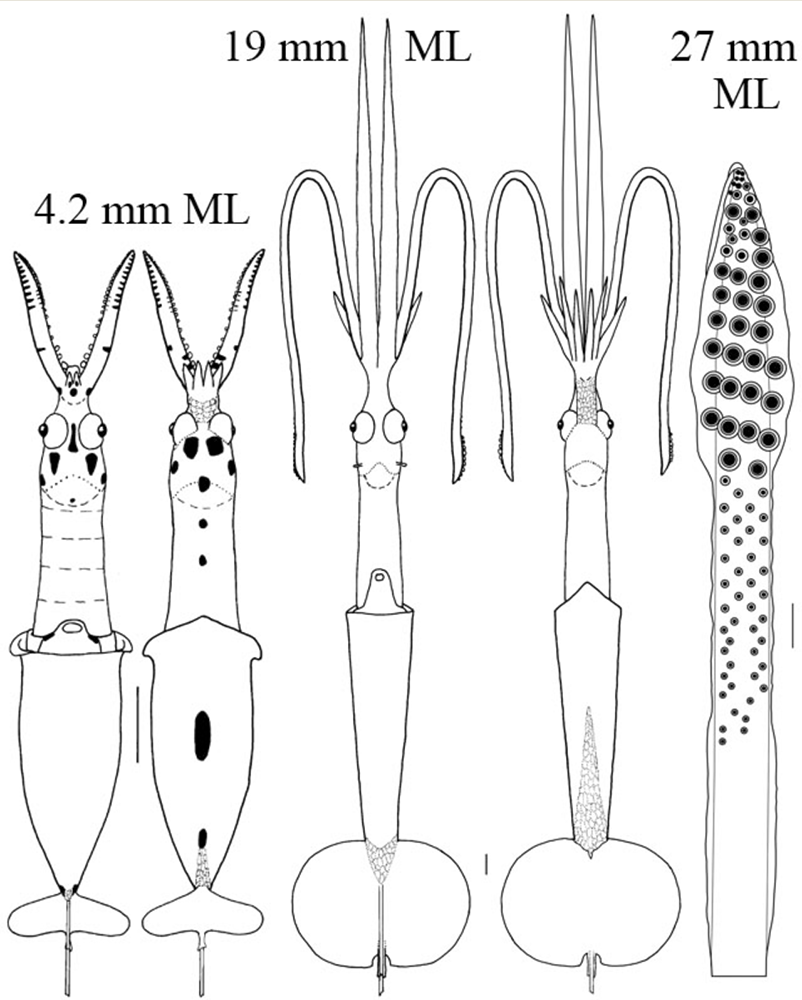 Figure. A. mangoldae paralarvae. Thumbnail (far left) - Ventral views showing relative sizes of the two paralarvae. Left - Ventral and dorsal views, 4.2 mm ML. Middle - Ventral and dorsal views, 19 mm ML. Right - Oral view of the tentacular club, 27 mm ML, with a typical primary doratopsid club (the distal expanded region with large suckers). The developing adult club is seen proximal to the doratopsid club on the tentacular stalk. This consists of a distal region with virtually no protective membranes and small suckers, and a proximal region with narrow protective membranes and no suckers. The developing adult club, therefore, shows the beginning of the features characteristic of the subadult. Scale bars = 1mm. Drawings from Young (1991).
Young, R. E. 1978. Vertical distribution and photosensitive vesicles of pelagic cephalopods from Hawaiian waters. Fish. Bull., 76: 583-615.
Young, R. E., M. Vecchione and C. F. E. Roper. 2007. A new genus and three new species of decapodiform cephalopods (Mollusca: Cephalopoda). Rev. Fish. Biol. Fisheries, 17: 353-365.